what type of cell acts to support nourish and regulate the development of sperm
Spermatozoa Evolution
From Embryology
Bound to:navigation, search
| Embryology - 8 April 2022 |
|---|
| Google Translate - select your language from the listing shown below (this will open a new external folio) |
| العربية | català | 中文 | 中國傳統的 | français | Deutsche | עִברִית | हिंदी | bahasa Republic of indonesia | italiano | 日本語 | 한국어 | မြန်မာ | Pilipino | Polskie | português | ਪੰਜਾਬੀ ਦੇ | Română | русский | Español | Swahili | Svensk | ไทย | Türkçe | اردو | ייִדיש | Tiếng Việt These external translations are automated and may not be accurate. (More? About Translations) |

Introduction
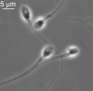
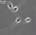
Human spermatozoa (light microscope)
Human spermatozoa (electron microscope)
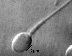
Single human spermatozoa[one]
This page introduces spermatogenesis the development of spermatozoa, the male haploid gamete cell. In humans at puberty, spermatozoa are produced by spermatogonia meiosis in the seminiferous tubules of the testis (male person gonad). A 2nd process of spermiogenesis leads to alter in cellular organisation and shape before release into the central lumen of the seminiferous tubule. This overall process has been variously divided into specific identifiable stages in different species: 6 in human, 12 in mouse, and 14 in rat. Structurally, the seminiferous tubule epithelium is divided into a basal and an upmost (adluminal) compartment by the claret–testis barrier (BTB). (More? Testis Development).
A second unique feature of this process is that during mitosis and meiosis the dividing cells remain connected by cytoplasmic bridges as the cells do non consummate cytokinesis. This cellular organization is described as a syncytium, only ending with release into the primal lumen of the seminiferous tubule, when the cell cytoplasm is discarded. Retinoic acid has been shown to be a cardinal regulator of the development procedure. (More? Retinoic acid)
- In a healthy adult homo male information technology takes about 48 days from meiosis to produce a mature spermatozoa, and he produces somewhere between 45 to 207 1000000 spermatozoa per twenty-four hours, or near 1 to 2,000 every 2nd. (More? Statistics)
| |

| Fertilization Links: fertilization | oocyte | spermatozoa | meiosis | | ovary | testis | menstrual cycle | zona pellucida | zygote | granulosa cell Lecture - Fertilization | 2016 Lecture | mitosis | Lecture - Week 1 and 2 | hydatidiform mole | Assisted Reproductive Engineering | | morula | blastocyst | Lecture - Genital Development | Category:Fertilization | ||
|
| Genital Links: genital | Lecture - Medicine | Lecture - Science | Lecture Movie | Medicine - Practical | primordial germ cell | meiosis | endocrine gonad | Genital Movies | genital abnormalities | Assisted Reproductive Technology | puberty | Category:Genital Female | 10 | X inactivation | ovary | corpus luteum | oocyte | uterus | vagina | reproductive cycles | menstrual cycle | Category:Female person Male | Y | SRY | testis | spermatozoa | ductus deferens | penis | prostate | Category:Male person | ||||
|
Medicine Practical | fertilization | Category:Spermatozoa
Some Recent Findings
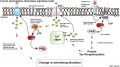
- Mechanism of Acrosome Biogenesis in Mammals [2] "During sexual reproduction, two haploid gametes fuse to class the zygote, and the acrosome is essential to this fusion process (fertilization) in animals. The acrosome is a special kind of organelle with a cap-like structure that covers the anterior portion of the caput of the spermatozoon. The acrosome is derived from the Golgi apparatus and contains digestive enzymes. With the progress of our understanding of acrosome biogenesis, a number of models have been proposed to address the origin of the acrosome. The acrosome has been regarded every bit a lysosome-related organelle, and information technology has been proposed to take originated from the lysosome or the autolysosome. Our review will provide a cursory historical overview and highlight recent findings on acrosome biogenesis in mammals."
- The dynamics and regulation of chromatin remodeling during spermiogenesis [iii] "The functional sperm is the key factor for species continuation. The process spermatogenesis, to produce mature sperm is quite complex. It begins with the proliferation and differentiation of spermatogonia, which develop from master spermatocytes to secondary spermatocytes and circular spermatids, which eventually develop into fertile mature sperm. Spermiogenesis is the latest stage of spermatogenesis, where the circular spermatids undergo a serial of dramatic morphological changes and farthermost condensation of chromatin to construct mature sperm with species-specific shape. During spermiogenesis, chromatin remodeling is a unique progress. It leads the nucleosome from a histone-based construction to a mostly protamine-based configuration. The main events of chromatin remodeling are the replacement of histone past histone variants, hyperacetylation, transient Deoxyribonucleic acid strand breaks and repair, variants by transition proteins and finally by protamines. In this review, we synthesize and summarize the current cognition on the progress of chromatin remodeling during spermiogenesis. We straighten out the chronological order of chromatin remodeling and illustrate the possible regulation mechanisms of each step."
- Deficiency of fibroblast growth factor ii (FGF-2) leads to abnormal spermatogenesis and contradistinct sperm physiology [iv] "In previous studies, we described the presence of fibroblast growth cistron ii (FGF-two) and its receptors (FGFRs) in human testis and sperm, which are involved in spermatogenesis and in motility regulation. The aim of the present study was to clarify the role of FGF-2 in the maintenance of sperm physiology using FGF-two knockout (KO) mice. Our results showed that in wild-type (WT) animals, FGF-2 is expressed in germ cells of the seminiferous epithelium, in epithelial cells of the epididymis, and in the flagellum and acrosomal region of epididymal sperm.... Overall, our findings advise that FGF-2 exerts a office in mammalian spermatogenesis and that the lack of FGF-2 leads to dysregulated sperm production and altered sperm morphology and office. FGF-2-deficient mice constitute a model for the study of the complex mechanisms underlying mammalian spermatogenesis."
- Small RNAs Are Trafficked from the Epididymis to Developing Mammalian Sperm [5] "The biogenesis of the RNA payload of mature sperm is of great interest, considering RNAs delivered to the zygote at fertilization can affect early evolution. Here, we tested the hypothesis that small RNAs are trafficked to mammalian sperm during the process of post-testicular maturation in the epididymis. By characterizing small RNA dynamics during germ cell maturation in mice, we confirm and extend prior observations that sperm undergo a dramatic switch in the RNA payload from piRNAs to tRNA fragments (tRFs) upon exiting the testis and entering the epididymis. Small RNA commitment to sperm could be recapitulated in vitro by incubating testicular spermatozoa with head epididymosomes. Finally, tissue-specific metabolic labeling of RNAs in intact mice definitively shows that mature sperm conduct RNAs that were originally synthesized in the epididymal epithelium. These data demonstrate that soma-germline RNA transfer occurs in male mammals, about likely via vesicular ship from the epididymis to maturing sperm."
- Review - Sperm nuclear protamines: A checkpoint to control sperm chromatin quality [vi] "Protamines are nuclear proteins which are specifically expressed in haploid male germ cells. Their replacement of histones and binding to Dna is followed by chromatin hypercondensation that protects DNA from negative influences past environmental factors. Mammalian sperm incorporate two types of protamines: PRM1 and PRM2. While the proportion of the two protamines is highly variable between unlike species, abnormal ratios within a species are known to exist associated with male person subfertility. Therefore, information technology is more than likely that right protamine expression represents a kind of chromatin checkpoint during sperm development rendering protamines as suitable biomarkers for the interpretation of sperm quality. This review presents an overview of our electric current knowledge on protamines comparing factor and poly peptide structures between different mammalian species with particular consideration given to man, mouse and stallion."
| More recent papers |
|---|
| This table allows an automated computer search of the external PubMed database using the listed "Search term" text link.
More? References | Word Page | Periodical Searches | 2019 References | 2020 References Search term: Spermatozoa Development | Spermatogonia Evolution | Spermatogonia Stem Cells | Spermatozoa Meiosis | Primary Spermatocyte | Secondary Spermatocyte | Spermatid | Spermatogenesis | Spermiogenesis | Sertoli Cell Evolution | Spermatozoa Chemotaxis | Acrosome |

| Older papers |
|---|
| These papers originally appeared in the Some Recent Findings tabular array, but as that list grew in length have now been shuffled down to this collapsible table. See likewise the Discussion Folio for other references listed by year and References on this current page.
|
Spermatozoa Movies
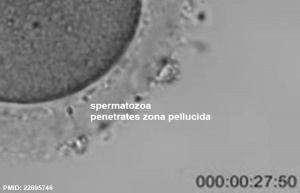
Human spermatozoa penetrating the zona pellucida during fertilisation (see Movie).
See as well Week 1 movies.
|
- Links: Week 1 movies | Movies
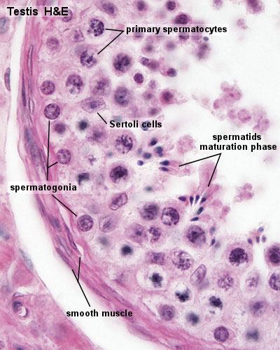
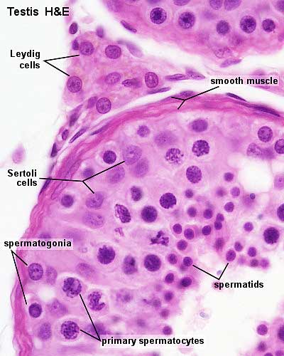
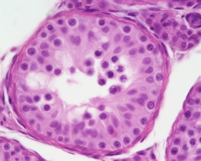
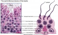
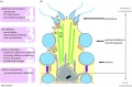
Seminiferous Tubule
| Adult Seminiferous tubule showing spermatozoa developmental stages | Seminiferous tubule cantankerous-section and supporting cells |
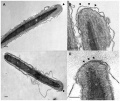
Rat Spermatogenesis drawing[12]
Seminiferous tubule cartoon [13]
- Spermatogonia - are the first cells of spermatogenesis
- Primary spermatocyte - large, enter the prophase of the outset meiotic division
- Secondary spermatocytes - small, complete the second meiotic division
- Spermatid - immature spermatozoa
- Spermatozoa - differentiated gamete
- Spermatozoa development: primordial germ cell - spermatogonia - primary spermatocyte - Template:Secondary spermatocyte - spermatid - spermatozoa
- Links: Testis Histology | testis
Spermatozoa Structure

Spermatozoa (mouse) cross-sections of tail (EM) and diagram[14]
-

Celebrated testis cartoon
-

Adult Seminiferous tubule showing spermatozoa developmental stages
-

Seminiferous tubule cantankerous-department and supporting cells
-
Human spermatozoon
-
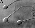
Human being spermatozoa
-

Single human spermatozoon
-
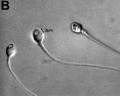
Man vacuolated spermatozoon
-
Celebrated EM spermatozoon tail
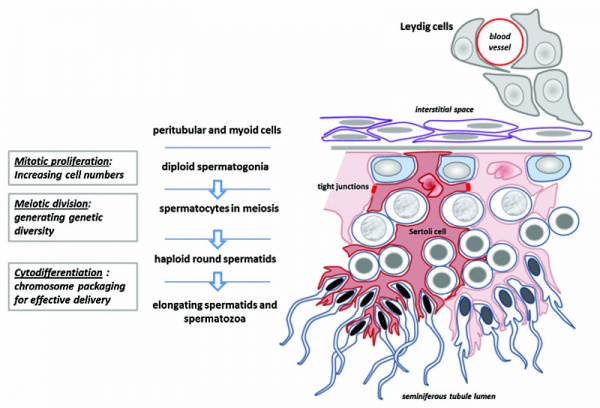
Other main cell types seen in the histological sections
- sertoli cells- support cells seen inside the seminiferous tubule
- Interstitial cells or Leydig cells - produce hormone
- Smooth musculus - surroundings seminiferous tubule and contribute to contraction of the tubule
Human Spermatozoa Development
- Spermatogenesis process of spermatagonia mature into spermatazoa (sperm).
- Continuously throughout life occurs in the seminiferous tubules in the male gonad- testis (plural testes).
- At puberty spermatagonia activate and proliferate (mitosis).
- about 48 days from inbound meiosis until morphologically mature spermatozoa
- about 64 days to consummate spermatogenesis, depending reproduction time of spermatogonia
- follicle stimulating hormone (FSH) - stimulates the spermatogenic epithelium
- luteinizing-hormone (LH) - stimulates testosterone production by Leydig cells
Spermatogonia
In humans at about 2 months of age, primordial germ cells (gonocytes) are replaced past adult dark (Ad) and pale (Ap) spermatogonia forming the spermatogonial stem jail cell (SSC) population that at puberty volition commence differentiation into spermatozoa.
The spermatogonia are the diploid stem cell (spermatogonial stem jail cell, SSC) progenitor for spermatozoa development. They are located on the basal lamina around the periphery of the seminiferous tubule wall. See this recent spermatogonia review.[xv]
In 1963 Clermont identified spermatogonia as Ap (pale) and Ad (dark) on basis of light microscope staining.[16]
- at present also blazon B
- sixty years - Ap spermatogonia number subtract
- 80 years - Ad spermatogonia number decrease
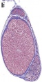
| Pre-puberty | Post-puberty |
|---|---|
 |  |
| Containing only spermatogonia and sertoli cells | Containing spermatogonia, sertoli cells and stages of spermatozoa jail cell meiosis |
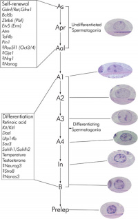 | Mouse spermatogonia have been shown to require a number of factors to regulate both their spermatogonial self-renewal and differentiation.Zhou Q & Griswold Dr.. (2008). Regulation of spermatogonia. , , . PMID: 20614596 DOI. The mouse "As model" originally stated that the As spermatogonia are the SSCs. A more recent proposal suggests that only some of the Adue south spermatogonia have the potential for long-term self-renewal, while others have a express capacity, indicating the presence of a SSC bureaucracy.[15] In the mouse testis, spermatogonial stalk cells can also be identified by Id4 expression[17], a ascendant-negative transcription factor containing a basic helix-loop-helix (bHLH) region. Id4 inhibits bounden to Dna and transcriptional transactivation by hetero-dimerization with other bHLH proteins. |
Meiosis
Spermatozoa maturation involves ii processes meiosis and spermiogenesis. Afterwards puberty, new spermatozoa continue to exist generated throughout life from a spermatogonia stem cell (SSC) population in the testis.
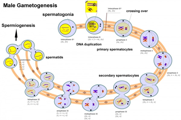
Primary Spermatocyte

The outset large differentiating diploid (2N) cell before meiosis I, that enters the prophase of the offset meiotic segmentation.
- Search PubMed: Primary Spermatocyte
Secondary Spermatocyte
The primary spermatocyte forms ii second pocket-sized cells that complete the second meiotic division to exist haploid (N) . Histologically difficult to detect, these cells will complete meiosis, forming adjacent the spermatid stage that is the immature morphological state of the spermatozoa.
- Search PubMed: Secondary Spermatocyte
Differences in Mammalian Meioses
| Female Oogenesis | Male Spermatogenesis | |
| Meiosis initiated | once in a finite population of cells | continuously in mitotically dividing stem jail cell population |
| Gametes produced | ane / meiosis | four / meiosis |
| Meiosis completed | delayed for months or years | completed in days or weeks |
| Meiosis Abort | arrest at 1st meiotic prophase | no arrest differentiation proceed continuously |
| Chromosome Equivalence | All chromosomes exhibit equivalent transcription and recombination during meiotic prophase | Sexual practice chromosomes excluded from recombination and transcription during outset meiotic prophase |
| Gamete Differentiation | occurs while diploid (in offset meiotic prophase) | occurs while haploid (later on meiosis ends) |
- Links: meiosis
Spermiogenesis
Spermiogenesis is the terminal phase of spermatogenesis, morphological changes transform the circular spermatids into the mature spermatozoa shape and structure.
- Nuclear pinch - chromatin condensation occurs by the replacement of histones with protamines.
- Acrosome formation - located over the anterior function of the spermatid nucleus, cap-like membrane-spring organelle formed through coalescence of the coated vesicles budding from the trans-Golgi network. The acrosome-acroplaxome-manchette circuitous is a major driver for the shaping of the spermatozoa head.
- Tail development - located over the posterior part of the spermatid nucleus, initially a centriole pair moves, the axoneme develops from the distal centriole. Axoneme consists of a primal pair of microtubules surrounded by 9 outer doublet microtubules ("nine × two + 2").
- Cytoplasm disposal - cytoplasm transported towards the tail along the manchette and finally its removal.
Other Features
- Nuclear pore redistribution - with packaging of the nuclear pores into the redundant and discarded nuclear envelope. [9]
- Autophagy - a self-digestion process, may likewise occur regulating cytoskeleton reorganization.[18] [19]
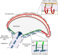
Sertoli Cell
The sertoli cell was named later Enrico Sertoli (1842 - 1910) an Italian (Milan) physiologist and histologist. These cells support spermatozoa development and span the wall of the seminiferous tubule.
- sustentacular cells of seminiferous tubules.
- form a "blood-testis" barrier through junctional complexes
- separate the intra-tubular germinal epithelium into 2 compartments
- basal compartment - cells are exposed to the actress-tubular environment
- luminal compartment - cells are discipline to an environment produced by Sertoli cells and germ cells
During infancy and childhood, sertoli cells are the about active prison cell population in the seminiferous tubule producing AMH from fetal catamenia until mid-puberty.[20]
- Links: sertoli cell | AMH
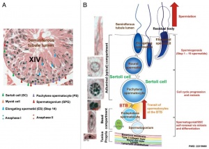
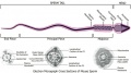
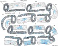
Spermatogenic Cycle
Along the length of the seminiferous tubule spermatozoa develop in a cyclic fashion over fourth dimension progressing through a number of stages, chosen the spermatogenic cycle, see review.[21] The number of stages appears to differ between species, in mouse there are 12 stages (I – XII) and in the rat fourteen stages.
In mouse, 1 spermatogenic cycle (12 stages) occurs over viii.vi days and four cycles (35 days) are required from spermatogonial stem prison cell to released spermatozoa.
Human being[22]
Gerbil[23]
Spermatozoa Construction

Spermatozoa (mouse) cross-sections of tail (EM) and diagram[fourteen]
Acroplaxome
This construction forms the acrosome plate with intermediate filament bundles of the marginal ring at the leading border of the acrosome. The acroplaxome site for Golgi-derived proacrosomal vesicles to tether and fuse and anchors the developing acrosome to the elongating spermatid head and may provide a scaffolding for the shaping of the spermatid nucleus.[24]
Acrosome
Derived from the Golgi appliance in conjunction with transient specialized bundles of microtubules (Template:Manchette), this vesicle releases its contents following progesterone stimulus or zona pellucida binding.
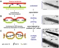
Acrosome Reaction
The "acrosome reaction" (AR) a type of specialised exocytosis, or like to the release of an exosome.[25] This also leads to changes in the spermatozoa membrane. The 26S proteasome[26], has been identified from in vitro studies to exist required for zona lysin in many species, including mammals.[27]
- Calcium ion permeability - Ca(2+) release from the acrosome leads to exocytosis of the acrosomal vesicle[28], alkalization appears to be a critical pace.[29]
- shop-operated Ca(2+) channels and voltage-dependent Ca(2+) channels
- Vesicle membrane - initially holding excreting molecules, remains on the cell surface
- Membrane loss - both outer acrosomal membrane and plasma membrane are lost by forming vesicles during acrosome reaction.
Acrosome reaction has a slow and rapid component:[30]
- Rapid - (seconds) efflux of calcium from intracellular stores, triggers fusion pores opening and the release of hybrid vesicles.
- Slow - (minutes) acrosomal swelling, triggered by activation of an adenylyl cyclase downstream of the opening of store-operated calcium channels. Determines the kinetics of the acrosome reaction.
- Exosomes - extracellular vesicles that are released from cells upon fusion of an intermediate endocytic compartment, the multivesicular body (MVB), with the plasma membrane.
- Proteasomes - protein complexes which typically degrade unneeded or damaged proteins by proteolysis.
Acrosin
- Acrosin is essential for sperm penetration through the zona pellucida in hamsters PNAS February 4, 2020 117 (5) 2513-2518 "Mammalian oocytes are surrounded by the zona pellucida, a glycoprotein coat that protects the oocyte and embryo from mechanical damage during their preimplantation evolution within the oviduct. Fertilizing spermatozoa must penetrate the zona, but we practice non know the exact mechanisms underlying this process. Sperm proteases were idea to work as zona lysins, but factor-knockout studies in mice did not back up this assumption. In this study, we generated hamsters without acrosin, the major acrosomal protease, to examine its role in both in vivo and in vitro fertilization. Surprisingly, mutant male hamsters were completely infertile because their spermatozoa were unable to penetrate the zona. We thus demonstrated that, at to the lowest degree in hamsters, acrosin is essential for sperm penetration through the zona."
Human acrosin - 22q13.33
- Links: OMIM - Acrosin
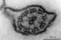
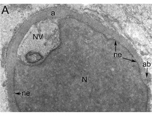
Nucleus
The spermatozoa nucleus undergoes all-encompassing pinch, and nuclear Deoxyribonucleic acid chromatin remodelling by tightly packing with spermatozoa-specific protamines.[31]
| Information technology is thought that the lysine-rich poly peptide precursor (H1 histone) has evolved into the arginine-rich protamines.[32] | Iii major spermatozoa nuclear basic proteins types:
|
| EM Human Spermatozoa Nucleus | |
|---|---|
 | 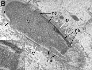 |
| Cap-stage spermatid nucleus[33] | Elongated spermatid nucleus[33] |
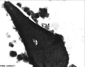 | 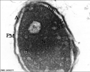 |
| Normal man spermatozoa[34] | Abnormal human spermatozoa[34] |
Axoneme
The stable mature microtubule-containing tail of the sperm.
Centriole
Spermatozoa initially contains ii centrioles (proximal, distal) and at fecundation only a single (proximal) is present, which in nearly mammalian species is contributed to reconstitute the zygotic centrosome. Note that in rodents (rat, mice) both centrioles are lost and only a maternal centrosomal inheritance occurs.
- distal centriole - (perpendicular to membrane) required as the basal body generating the microtubule axoneme and is and so lost (disintegration).
- proximal centriole - required after fertilisation for decondensing spermatozoa nucleus allowing development into the male person pronucleus.
Manchette
A transient microtubule structure formed in spermatids involved in the process of: associates of the mammalian sperm tail, mechanical shaping and condensation of the sperm nucleus. These microtubules are also invloved with specific transport, intramanchette ship, which has been likened to intraflagellar transport. This microtubular structure surrounds the nucleus of the developing spermatid and is thought too to assist in both the reshaping of the nucleus and redistribution of spermatid cytoplasm.
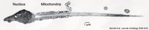
Mitochondria
Independent in the initial segment provide the energy for motility and may also enter the egg on fertilization, but are eliminated past a ubiquitin-dependent machinery.
Perinuclear Theca
Located in the sperm head perinuclear region and contains a cytoskeletal element to maintain the shape of the sperm head and functional molecules leading to oocyte activation during fertilization.
Mature Human Spermatozoa
| | Features:
Human Spermatazoa Statistics | Development Animation - Spermatozoa |
Human Spermatozoa Statistics
| Spermatozoa | Number/Fourth dimension |
|---|---|
| Production | |
| produced / day (2 testes) | 45 to 207 meg |
| compare adult human red blood cell / twenty-four hours | 250,000 million |
| produced / second each day (approx) | 2,000 |
| Storage | |
| stored (epididymal reserves) up to per epididymis | 182 one thousand thousand |
| stored extragonadal | 440 million |
| extragonadal - ductuli deferentia and caudae epididymides per ejaculation | 225 million |
| Transit Times | |
| through the head | 0.72 mean solar day |
| through the corpus | 0.71 days |
| through the cauda epididymidis | 1.76 days |
| Tabular array Data [35] See also WHO human semen reference values(2010).[36] Links: spermatozoa | |
Spermatozoa Morphology
Morphology is a term used to describe the overall appearance of a cell or tissue and is often used to characterise changes in cellular country or activity. Historically, in that location have been studies comparison the overall appearance of spermatozoa betwixt different species.[37] More than recently, there accept been several different means of characterising the morphology of human spermatozoa developed mainly in relation to clinical reproductive technologies.
Integrated Sperm Analysis System (ISAS)
A semi-automated computer-aided system that measures spermatozoa head parameters length (L), width (W), area (A), perimeter (P), acrosomal area (Ac), and the derived values L/Westward and P/A.[38]
- For each man a homogeneous population of distributions characterized seminal spermatozoa (seven,942 cells: median values 50 4.4 μm, W ii.viii μm, A 9.8 μm(two), P 12.5 μm, Ac 47.5%, Fifty/W 1.57, P/A 1.27)
- Unlike men could have spermatozoa of significantly different dimensions.
- Head dimensions for swim-up spermatozoa from dissimilar men (4 812 cells) were similar to those in semen, differing only by 2%-five%.
- The values of 50, Westward and 50/W fell within the limits given past the World Health Organization (WHO).
- A subpopulation of 404 spermatozoa considered to fit the stringent criteria of WHO 'normal' seminal spermatozoa[36] from both semen and swim-up were characterized by median values (and 95% conviction intervals) of 50, 4.3 μm (3.8-4.9), W, 2.ix μm (2.6-3.three), A, 10.2 μm(2) (viii.five-12.2), P, 12.iv μm (eleven.iii-13.ix), Ac, 49% (36-60), 50/Due west, 1.49 (ane.32-1.67) and P/A, ane.22 (1.eleven-ane.35). These median values autumn within the 95th centile confidence limits given past WHO, simply the confidence intervals for Fifty and Due west were larger.
Spermatozoa Chemotaxis
Chemotaxis was outset identified in marine species[39], which nonetheless remains today as a model system. While the signals may differ, the overall effect is to chemically assault spermatozoa to the oocyte to allow fertilisation to occur.
The following series of 2011 enquiry articles have identified the spermatozoa calcium channel protein (CatSper) as the progesterone activated pathway involved in capacitated spermatozoa chemotaxis.
|

Human Spermatozoa Chemotaxis Model (2009)[43]
See also 2008 review.[44]
Sertoli Cell
The sertoli cells are the first cells to exist differentiated in development past SRY expression. Post-puberty these are the "back up" cells for spermatozoa development and ship from the periphery to lumen of the seminiferous tubule. Sertoli cells form a barrier with cell junctions at the Sertoli cell-jail cell and Sertoli-germ cell interface.
Sertoli jail cell postnatal proliferation may be regulated by thyroid status. An fauna model study of postnatal transient hypothyroidism has demonstrated Sertoli cell proliferation (6 to 8 fold increase) 2 days after the diet switch and remained elevated the next days.[45]
Histology
-
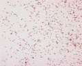
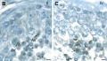
Homo spermatozoa, x20, Papanicolaou stain
-
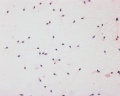
Human spermatozoa, x40, Papanicolaou stain
-
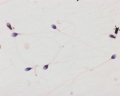
Human spermatozoa, x100, Papanicolaou stain
Papanicolaou stain (Papanicolaou'south stain, Pap stain) a multichromatic (v dyes) staining histological technique developed by George Papanikolaou, used to differentiate cells in smear preparations of diverse bodily secretions.
- Links: sertoli jail cell | Testis Histology | Histology Stains | Search PubMed - Sertoli Cell Evolution
Male person Abnormalities
Male Infertility Genes
Examples of known genes resulting in various forms of male infertility.
| Gene abbreviation | Name | Gene Location | Online Mendelian Inheritance in Man (OMIM) | HUGO Gene Classification Committee (HGNC) | GeneCards (GCID) | Diagnosis |
|---|---|---|---|---|---|---|
| AURKC | Aurora kinase C | 19q13.43 | 603495 | 11391 | GC19P057230 | Macrozoospermia |
| CATSPER1 | Cation channel sperm-associated ane | 11q13.1 | 606389 | 17116 | GC11M066034 | Asthenozoospermia |
| CFTR | Cystic fibrosis transmembrane conductance regulator | sevenq31.2 | 602421 | 1884 | GC07P117465 | Obstructive azoospermia |
| DNAH1 | Dynein axonemal heavy chain one | 3p21.1 | 603332 | 2940 | GC03P052350 | Asthenozoospermia |
| DPY19L2 | Dpy-nineteen-like ii gene | 12q14.2 | 613893 | 19414 | GC12M063558 | Globozoospermia |
| GALNTL5 | Polypeptide Northward-acetylgalactosaminyltransferase-similar v | 7q36.1 | 615133 | 21725 | GC07P151956 | Asthenozoospermia |
| MAGEB4 | MAGE family member B4 | Xp21.ii | 300153 | 6811 | GC0XP030260 | Azoospermia |
| NANOS1 | Nanos C2HC-type zinc finger i | 10q26.11 | 608226 | 23044 | GC10P119029 | Azoospermia |
| NR0B1 | Nuclear receptor subfamily 0 group B member 1 | Xp21.ii | 300473 | 7960 | GC0XM030322 | Azoospermia |
| NR5A1 | Nuclear receptor subfamily 5 grouping A member 1 | nineq33.3 | 184757 | 7983 | GC09M124481 | Azoospermia |
| SOHLH1 | Spermatogenesis and oogenesis-specific basic helix–loop–helix i | 9q34.3 | 610224 | 27845 | C09M135693 | Azoospermia |
| vSPATA16 | Spermatogenesis-associated xvi | 3q26.31 | 609856 | 29935 | GC03M172889 | Globozoospermia |
| SYCE1 | Synaptonemal complex central element protein 1 | 10q26.3 | 611486 | 28852 | GC10M133553 | Azoospermia |
| TAF4B | TATA-box binding protein-associated factor 4b | xviiiq11.2 | 601689 | 11538 | GC18P026225 | Azoospermia |
| TEX11 | Testis expressed 11 | Tenq13.1 | 300311 | 11733 | GC0XM070528 | Azoospermia |
| TEX15 | Testis expressed 15, meiosis and synapsis associated | 8p12 | 605795 | 11738 | GC08M030808 | Azoospermia |
| WT1 | Wilms tumour 1 | eightp12 | 607102 | 12796 | GC11M032365 | Azoospermia |
| ZMYND15 | Zinc-finger MYND-blazon containing 15 | 17p13.two | 614312 | 20997 | GC17P004740 | Azoospermia |
| Tabular array data source[46] (tabular array 1)Links: fertilization | spermatozoa | testis | Male Infertility Genes | Female Infertility Genes | oocyte | ovary | Genetic Abnormalities | ART Asthenozoospermia - (asthenospermia) term for reduced spermatozoa motility. Azoospermia - term for no spermatozoa located in the ejaculate. Globozoospermia - term for spermatozoa with a circular head and no acrosome. | ||||||
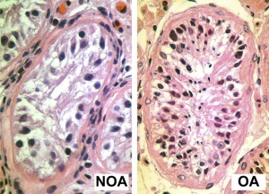
Human Seminiferious Tubule - Not-obstructive azoospermia and Obstructive azoospermia
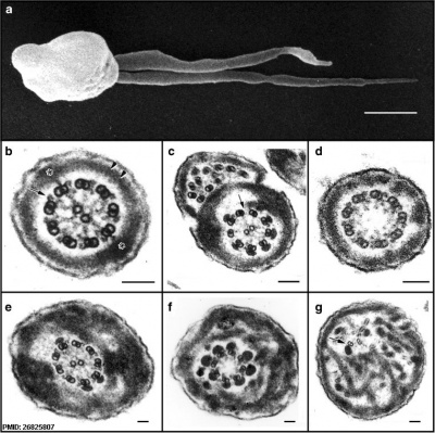
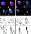
Homo sperm pathologies[47] These electron micrographs show a range of tail structural abnormalities including (a) two tails. (b) shows a normal spermatozoa tail cross-section and c to 1000 testify a range of abnormal tail structures (open paradigm to see details).
Johnsen score
A clinical score (1-x) for assessing spermatogenesis in a human testicular biopsy. Named afterwards the writer of the original commodity .[48]
| Johnsen score | Description |
|---|---|
| 10 | complete spermatogenesis and perfect tubules |
| 9 | many spermatozoa present but disorganized spermatogenesis |
| 8 | just a few spermatozoa present |
| 7 | no spermatozoa but many spermatids present |
| half-dozen | only a few spermatids present |
| v | no spermatozoa or spermatids present but many spermatocytes present |
| 4 | only a few spermatocytes nowadays |
| 3 | just spermatogonia present |
| 2 | no germ cells present |
| 1 | neither germ cells nor Sertoli cells present |
| Reference: Johnsen SG. Testicular biopsy score count - a method for registration of spermatogenesis in human testes: normal values and results in 335 hypogonadal males. (1970) Hormones 1(1): two-25. PubMed 5527187 | |
| Classification | Count (Millions/mL) |
|---|---|
| Azoospermia | 0 |
| Severe oligozoospermia | less than 1 |
| Moderate oligozoospermia | 1-5 |
| Mild oligozoospermia | 5-20 |
| Normal | greater than 20 |
Oligospermia
(Low Sperm Count) less than 20 million sperm after 72 hour abstinence from sex
Azoospermia
(Absent Sperm) blockage of duct network
Immotile Cilia Syndrome
Lack of sperm movement
Acephalic spermatozoa syndrome
Acephalic spermatozoa syndrome is characterized by the presence of very few intact spermatozoa and tailless sperm heads in the semen and leads to astringent male infertility. Sad1 and UNC84 domain-containing 5 (SUN5) is a testis-specific nuclear envelope protein. A recent study has shown that mutations in SUN5 announced to touch on the secondary structure of the protein and influence its folding and cellular localization.[49]
- Links: OMIM - SUN5
Infertility - Stem Cells
Recent studies accept been able to transplant of own cryostored spermatogonial stem cells (SSCs) is a promising technique for fertility restoration when the SSC pool has been depleted.[fifty]
Additional Images
-
Testis histology
-
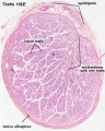
Testis histology, young and mature - H&Due east
-
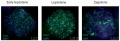
Human testis NANOG expression
-
Man spermatozoa acrosomal poly peptide SP-x[33]
-
Homo spermatid electron micrograph[33]
-
Model capacitation-induced acrosome docking to sperm membrane[xiv]
-
Mouse spermiogenesis model[14]
-
Mouse- seminiferous tubule histology[14]
-
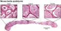
Mouse- epididymis histology
-
Mouse- spermatozoa EM and diagram
-
Mouse - Spatiotemporal progression of annulus during mouse spermiogenesis
-
Mouse spermatogonia meiotic prophase I stages
-

Rat Spermatogenesis figure[51]
-
EM - Capacitation alters the ultrastructure of the apical head and the acrosome of boar sperm
-
Human spermatozoa - phospholipase C zeta localization[52]
-
Chemotaxis Model[43]
-
Labeled Chemotaxis Model[43]
-
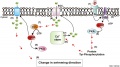
Spermatogenesis androgen action[53]
-
Mouse spermatogenesis stages[54]
References
- ↑ Oliveira JB, Petersen CG, Massaro FC, Baruffi RL, Mauri AL, Silva LF, Ricci J & Franco JG. (2010). Motile sperm organelle morphology examination (MSOME): intervariation study of normal sperm and sperm with large nuclear vacuoles. Reprod. Biol. Endocrinol. , 8, 56. PMID: 20529256 DOI.
- ↑ Khawar MB, Gao H & Li W. (2019). Mechanism of Acrosome Biogenesis in Mammals. Forepart Cell Dev Biol , vii, 195. PMID: 31620437 DOI.
- ↑ Hao SL, Ni FD & Yang WX. (2019). The dynamics and regulation of chromatin remodeling during spermiogenesis. Gene , 706, 201-210. PMID: 31085275 DOI.
- ↑ Saucedo L, Rumpel R, Sobarzo C, Schreiner D, Brandes G, Lustig L, Vazquez-Levin MH, Grothe C & Marín-Briggiler C. (2018). Deficiency of fibroblast growth factor 2 (FGF-2) leads to abnormal spermatogenesis and altered sperm physiology. J. Prison cell. Physiol. , , . PMID: 30054911 DOI.
- ↑ Sharma U, Sunday F, Conine CC, Reichholf B, Kukreja S, Herzog VA, Ameres SL & Rando OJ. (2018). Small-scale RNAs Are Trafficked from the Epididymis to Developing Mammalian Sperm. Dev. Jail cell , , . PMID: 30057273 DOI.
- ↑ Steger K & Balhorn R. (2018). Sperm nuclear protamines: A checkpoint to control sperm chromatin quality. Anat Histol Embryol , 47, 273-279. PMID: 29797354 DOI.
- ↑ Levine H, Jørgensen N, Martino-Andrade A, Mendiola J, Weksler-Derri D, Mindlis I, Pinotti R & Swan SH. (2017). Temporal trends in sperm count: a systematic review and meta-regression assay. Hum. Reprod. Update , 23, 646-659. PMID: 28981654 DOI.
- ↑ Maree L, du Plessis SS, Menkveld R & van der Horst G. (2010). Morphometric dimensions of the human sperm head depend on the staining method used. Hum. Reprod. , 25, 1369-82. PMID: 20400771 DOI.
- ↑ 9.0 9.1 Ho HC. (2010). Redistribution of nuclear pores during formation of the redundant nuclear envelope in mouse spermatids. J. Anat. , 216, 525-32. PMID: 20136667 DOI.
- ↑ Bastián Y, Roa-Espitia AL, Mújica A & Hernández-González EO. (2010). Calpain modulates capacitation and acrosome reaction through cleavage of the spectrin cytoskeleton. Reproduction , 140, 673-84. PMID: 20716611 DOI.
- ↑ Pellegrini G, Di Siena South, Claps G, Di Cesare S, Dolci Southward, Rossi P, Geremia R & Grimaldi P. (2010). Microgravity promotes differentiation and meiotic entry of postnatal mouse male person germ cells. PLoS ONE , 5, e9064. PMID: 20140225 DOI.
- ↑ Cheng YH, Wong EW & Cheng CY. (2011). Cancer/testis (CT) antigens, carcinogenesis and spermatogenesis. Spermatogenesis , one, 209-220. PMID: 22319669 DOI.
- ↑ Hunter D, Anand-Ivell R, Danner S & Ivell R. (2012). Models of in vitro spermatogenesis. Spermatogenesis , 2, 32-43. PMID: 22553488 DOI.
- ↑ 14.0 xiv.one 14.2 14.iii 14.four xiv.five Borg CL, Wolski KM, Gibbs GM & O'Bryan MK. (2010). Phenotyping male infertility in the mouse: how to get the most out of a 'non-performer'. Hum. Reprod. Update , 16, 205-24. PMID: 19758979 DOI.
- ↑ 15.0 fifteen.1 de Rooij DG. (2017). The nature and dynamics of spermatogonial stem cells. Development , 144, 3022-3030. PMID: 28851723 DOI.
- ↑ Clermont Y. (1966). Spermatogenesis in man. A report of the spermatogonial population. Fertil. Steril. , 17, 705-21. PMID: 5920556
- ↑ Dominicus F, Xu Q, Zhao D & Degui Chen C. (2015). Id4 Marks Spermatogonial Stalk Cells in the Mouse Testis. Sci Rep , v, 17594. PMID: 26621350 DOI.
- ↑ Shang Y, Wang H, Jia P, Zhao H, Liu C, Liu West, Song Z, Xu Z, Yang L, Wang Y & Li West. (2016). Autophagy regulates spermatid differentiation via degradation of PDLIM1. Autophagy , 12, 1575-92. PMID: 27310465 DOI.
- ↑ Ozturk Due north, Steger G & Schagdarsurengin U. (2017). The affect of autophagy in spermiogenesis. Asian J. Androl. , nineteen, 617-618. PMID: 27905325 DOI.
- ↑ Grinspon RP & Rey RA. (2011). New perspectives in the diagnosis of pediatric male hypogonadism: the importance of AMH as a Sertoli cell marking. Arq Bras Endocrinol Metabol , 55, 512-9. PMID: 22218431
- ↑ Clermont Y. (1972). Kinetics of spermatogenesis in mammals: seminiferous epithelium bicycle and spermatogonial renewal. Physiol. Rev. , 52, 198-236. PMID: 4621362 DOI.
- ↑ Chaturvedi PK & Johnson 50. (1993). Architectural arrangement of stages of the spermatogenic cycle within homo seminiferous tubules is related to efficiency of spermatogenesis. Jail cell Tissue Res. , 273, 65-70. PMID: 8364962
- ↑ Naylor GJ, McNamee HB & Moody JP. (1970). The plasma control of erythrocyte sodium and potassium metabolism in depressive illness. J Psychosom Res , xiv, 179-86. PMID: 5477358
- ↑ Kierszenbaum AL & Tres LL. (2004). The acrosome-acroplaxome-manchette circuitous and the shaping of the spermatid caput. Arch. Histol. Cytol. , 67, 271-84. PMID: 15700535
- ↑ Okabe M. (2016). The Acrosome Reaction: A Historical Perspective. Adv Anat Embryol Cell Biol , 220, 1-13. PMID: 27194347 DOI.
- ↑ Bard JAM, Goodall EA, Greene ER, Jonsson East, Dong KC & Martin A. (2018). Structure and Role of the 26S Proteasome. Annu. Rev. Biochem. , 87, 697-724. PMID: 29652515 DOI.
- ↑ Zimmerman SW, Manandhar G, Yi YJ, Gupta SK, Sutovsky Thou, Odhiambo JF, Powell MD, Miller DJ & Sutovsky P. (2011). Sperm proteasomes degrade sperm receptor on the egg zona pellucida during mammalian fertilization. PLoS One , 6, e17256. PMID: 21383844 DOI.
- ↑ Beltrán C, Treviño CL, Mata-Martínez E, Chávez JC, Sánchez-Cárdenas C, Baker Thou & Darszon A. (2016). Office of Ion Channels in the Sperm Acrosome Reaction. Adv Anat Embryol Cell Biol , 220, 35-69. PMID: 27194349 DOI.
- ↑ Chávez JC, De la Vega-Beltrán JL, José O, Torres P, Nishigaki T, Treviño CL & Darszon A. (2018). Acrosomal alkalization triggers Ca2+ release and acrosome reaction in mammalian spermatozoa. J. Jail cell. Physiol. , 233, 4735-4747. PMID: 29135027 DOI.
- ↑ Sosa CM, Pavarotti MA, Zanetti MN, Zoppino FC, De Blas GA & Mayorga LS. (2015). Kinetics of human sperm acrosomal exocytosis. Mol. Hum. Reprod. , 21, 244-54. PMID: 25452326 DOI.
- ↑ Rathke C, Baarends WM, Awe S & Renkawitz-Pohl R. (2014). Chromatin dynamics during spermiogenesis. Biochim. Biophys. Acta , 1839, 155-68. PMID: 24091090 DOI.
- ↑ Saperas North & Ausió J. (2013). Sperm nuclear basic proteins of tunicates and the origin of protamines. Biol. Balderdash. , 224, 127-36. PMID: 23995738 DOI.
- ↑ 33.0 33.ane 33.ii 33.3 Westbrook VA, Schoppee PD, Vanage GR, Klotz KL, Diekman AB, Flickinger CJ, Coppola MA & Herr JC. (2006). Hominoid-specific SPANXA/D genes demonstrate differential expression in individuals and protein localization to a singled-out nuclear envelope domain during spermatid morphogenesis. Mol. Hum. Reprod. , 12, 703-16. PMID: 17012309 DOI.
- ↑ 34.0 34.1 Iranpour FG. (2014). The effects of protamine deficiency on ultrastructure of human sperm nucleus. Adv Biomed Res , 3, 24. PMID: 24592371 DOI.
- ↑ Amann RP & Howards SS. (1980). Daily spermatozoal product and epididymal spermatozoal reserves of the human male. J. Urol. , 124, 211-5. PMID: 6772801
- ↑ 36.0 36.1 Cooper TG, Noonan Due east, von Eckardstein S, Auger J, Baker HW, Behre HM, Haugen TB, Kruger T, Wang C, Mbizvo MT & Vogelsong KM. (2010). Globe Health System reference values for human semen characteristics. Hum. Reprod. Update , 16, 231-45. PMID: 19934213 DOI.
- ↑ Fawcett DW. (1970). A comparative view of sperm ultrastructure. Biol. Reprod. , 2, Suppl 2:90-127. PMID: 5521054
- ↑ Bellastella Grand, Cooper TG, Battaglia M, Ströse A, Torres I, Hellenkemper B, Soler C & Sinisi AA. (2010). Dimensions of human being ejaculated spermatozoa in Papanicolaou-stained seminal and swim-up smears obtained from the Integrated Semen Analysis System (ISAS(®)). Asian J. Androl. , 12, 871-ix. PMID: 20852650 DOI.
- ↑ Lillie FR. (1912). THE PRODUCTION OF SPERM ISO-AGGLUTININS Past OVA. Science , 36, 527-30. PMID: 17735765 DOI.
- ↑ Strünker T, Goodwin N, Brenker C, Kashikar ND, Weyand I, Seifert R & Kaupp UB. (2011). The CatSper channel mediates progesterone-induced Ca2+ influx in human sperm. Nature , 471, 382-6. PMID: 21412338 DOI.
- ↑ Johannessen JV. (1992). [Physicians and leadership]. Tidsskr. Nor. Laegeforen. , 112, 2950. PMID: 1412339
- ↑ Armon Fifty & Eisenbach M. (2011). Behavioral mechanism during human sperm chemotaxis: involvement of hyperactivation. PLoS ONE , 6, e28359. PMID: 22163296 DOI.
- ↑ 43.0 43.1 43.2 Teves ME, Guidobaldi HA, Uñates DR, Sanchez R, Miska West, Publicover SJ, Morales Garcia AA & Giojalas LC. (2009). Molecular mechanism for human sperm chemotaxis mediated by progesterone. PLoS ONE , 4, e8211. PMID: 19997608 DOI.
- ↑ Kaupp UB, Kashikar ND & Weyand I. (2008). Mechanisms of sperm chemotaxis. Annu. Rev. Physiol. , 70, 93-117. PMID: 17988206 DOI.
- ↑ Rijntjes Due east, Gomes MLM, Zupanič N, Swarts HJM, Keijer J & Teerds KJ. (2017). Transient Hypothyroidism: Dual Consequence on Adult-Blazon Leydig Cell and Sertoli Cell Development. Front Physiol , viii, 323. PMID: 28588502 DOI.
- ↑ Harper JC, Aittomäki Chiliad, Borry P, Cornel MC, de Wert Thou, Dondorp W, Geraedts J, Gianaroli L, Ketterson Yard, Liebaers I, Lundin K, Mertes H, Morris M, Pennings G, Sermon K, Spits C, Soini Southward, van Montfoort APA, Veiga A, Vermeesch JR, Viville S & Macek M. (2018). Recent developments in genetics and medically assisted reproduction: from research to clinical applications. Eur. J. Hum. Genet. , 26, 12-33. PMID: 29199274 DOI.
- ↑ Linck RW, Chemes H & Albertini DF. (2016). The axoneme: the propulsive engine of spermatozoa and cilia and associated ciliopathies leading to infertility. J. Assistance. Reprod. Genet. , 33, 141-56. PMID: 26825807 DOI.
- ↑ Johnsen SG. (1970). Testicular biopsy score count--a method for registration of spermatogenesis in human testes: normal values and results in 335 hypogonadal males. Hormones , 1, 2-25. PMID: 5527187
- ↑ Shang Y, Yan J, Tang West, Liu C, Xiao Due south, Guo Y, Yuan Fifty, Chen L, Jiang H, Guo X, Qiao J & Li West. (2018). Mechanistic insights into acephalic spermatozoa syndrome-associated mutations in the man SUN5 cistron. J. Biol. Chem. , 293, 2395-2407. PMID: 29298896 DOI.
- ↑ Kanbar Yard, de Michele F & Wyns C. (2018). Cryostorage of testicular tissue and retransplantation of spermatogonial stem cells in the infertile male person. Best Pract. Res. Clin. Endocrinol. Metab. , , . PMID: 30448111 DOI.
- ↑ Winawer SJ, Flehinger BJ, Buchalter J, Herbert Eastward & Shike Thou. (1990). Declining serum cholesterol levels prior to diagnosis of colon cancer. A time-trend, case-control study. JAMA , 263, 2083-5. PMID: 2319669
- ↑ Aarabi Grand, Yu Y, Xu W, Tse MY, Pang SC, Yi YJ, Sutovsky P & Oko R. (2012). The testicular and epididymal expression profile of PLCζ in mouse and human does non support its role as a sperm-borne oocyte activating cistron. PLoS ONE , 7, e33496. PMID: 22428063 DOI.
- ↑ Verhoeven 1000, Willems A, Denolet E, Swinnen JV & De Gendt K. (2010). Androgens and spermatogenesis: lessons from transgenic mouse models. Philos. Trans. R. Soc. Lond., B, Biol. Sci. , 365, 1537-56. PMID: 20403868 DOI.
- ↑ Phillips BT, Gassei M & Orwig KE. (2010). Spermatogonial stem cell regulation and spermatogenesis. Philos. Trans. R. Soc. Lond., B, Biol. Sci. , 365, 1663-78. PMID: 20403877 DOI.
Journals
- Spermatogenesis | PubMed - Spermatogenesis "Spermatogenesis is a new quarterly, peer-reviewed journal that will publish loftier-quality articles roofing all aspects of spermatogenesis." Note concluding PubMed entries for this periodical in 2016.
- WHO. WHO Laboratory Manual for the Exam and Processing of Human Semen. 5th ed. Geneva, Switzerland: World Health Organization; 2010. Online PDF
Reviews
de Rooij DG. (2017). The nature and dynamics of spermatogonial stem cells. Development , 144, 3022-3030. PMID: 28851723 DOI.
Griswold MD. (2016). Spermatogenesis: The Commitment to Meiosis. Physiol. Rev. , 96, 1-17. PMID: 26537427 DOI.
Talwar P & Hayatnagarkar Southward. (2015). Sperm part test. J Hum Reprod Sci , viii, 61-nine. PMID: 26157295 DOI.
Yoshida S. (2010). Stalk cells in mammalian spermatogenesis. Dev. Growth Differ. , 52, 311-vii. PMID: 20388168 DOI.
Hogarth CA & Griswold Md. (2010). The key function of vitamin A in spermatogenesis. J. Clin. Invest. , 120, 956-62. PMID: 20364093 DOI.
Ruwanpura SM, McLachlan RI & Meachem SJ. (2010). Hormonal regulation of male person germ cell development. J. Endocrinol. , 205, 117-31. PMID: 20144980 DOI.
Hermo 50, Pelletier RM, Cyr DG & Smith CE. (2010). Surfing the wave, bike, life history, and genes/proteins expressed past testicular germ cells. Role i: groundwork to spermatogenesis, spermatogonia, and spermatocytes. Microsc. Res. Tech. , 73, 241-78. PMID: 19941293 DOI.
Hermo L, Pelletier RM, Cyr DG & Smith CE. (2010). Surfing the wave, cycle, life history, and genes/proteins expressed past testicular germ cells. Part 2: changes in spermatid organelles associated with development of spermatozoa. Microsc. Res. Tech. , 73, 279-319. PMID: 19941292 DOI.
Kaupp UB, Kashikar ND & Weyand I. (2008). Mechanisms of sperm chemotaxis. Annu. Rev. Physiol. , lxx, 93-117. PMID: 17988206 DOI.
Eddy EM, Toshimori Chiliad & O'Brien DA. (2003). Fibrous sheath of mammalian spermatozoa. Microsc. Res. Tech. , 61, 103-15. PMID: 12672126 DOI.
de Rooij DG & Russell LD. (2000). All you wanted to know about spermatogonia but were afraid to ask. J. Androl. , 21, 776-98. PMID: 11105904
Clermont Y. (1972). Kinetics of spermatogenesis in mammals: seminiferous epithelium cycle and spermatogonial renewal. Physiol. Rev. , 52, 198-236. PMID: 4621362 DOI.
Articles
Cooper TG, Noonan East, von Eckardstein Due south, Auger J, Baker HW, Behre HM, Haugen TB, Kruger T, Wang C, Mbizvo MT & Vogelsong KM. (2010). World Health Organization reference values for homo semen characteristics. Hum. Reprod. Update , 16, 231-45. PMID: 19934213 DOI.
LEBLOND CP & CLERMONT Y. (1952). Definition of the stages of the wheel of the seminiferous epithelium in the rat. Ann. N. Y. Acad. Sci. , 55, 548-73. PMID: 13139144
NCBI Bookshelf
- StemBook [Internet]. Cambridge (MA): Harvard Stem Cell Institute; 2008 Regulation of spermatogonia
MBoC - Sperm | MBoC - Highly simplified drawing of a cross-section of a seminiferous tubule in a mammalian testis | MBoC - Cytoplasmic bridges in developing sperm cells and their precursors
- NCBI Bookshelf spermatozoa | spermatogenesis | spermiogenesis
Search
- Pubmed spermatozoa | spermatogenesis | spermiogenesis
Terms
| Spermatozoa Evolution (expand to run across terms) | ||
|---|---|---|
| Spermatozoa Development Notation there are additional glossaries associated with genital, spermatozoa, oocyte and renal. Spermatozoon
See also: Spermatozoa Terms collapse table
|

External Links
External Links Notice - The dynamic nature of the internet may hateful that some of these listed links may no longer function. If the link no longer works search the web with the link text or name. Links to any external commercial sites are provided for data purposes simply and should never exist considered an endorsement. UNSW Embryology is provided as an educational resource with no clinical information or commercial affiliation.
- World Health Arrangement - WHO Laboratory Transmission for the Examination and Processing of Human Semen. fifth ed. Geneva, Switzerland: World Health Organization; 2010. Online PDF
Glossary Links
- Glossary: A | B | C | D | East | F | G | H | I | J | Thou | L | Thousand | N | O | P | Q | R | S | T | U | V | Due west | X | Y | Z | Numbers | Symbols | Term Link
Cite this page: Hill, M.A. (2022, April viii) Embryology Spermatozoa Evolution. Retrieved from https://embryology.med.unsw.edu.au/embryology/index.php/Spermatozoa_Development
-
- What Links Here?
- © Dr Marking Hill 2022, UNSW Embryology ISBN: 978 0 7334 2609 4 - UNSW CRICOS Provider Code No. 00098G
Source: https://embryology.med.unsw.edu.au/embryology/index.php/Spermatozoa_Development
0 Response to "what type of cell acts to support nourish and regulate the development of sperm"
Post a Comment